Post-orthodontic rehabilitation
Finished your braces, but the shape of your teeth, the gaps left or the way they meet are not what you expected? This page explains why that happens and what the options are.
On this page
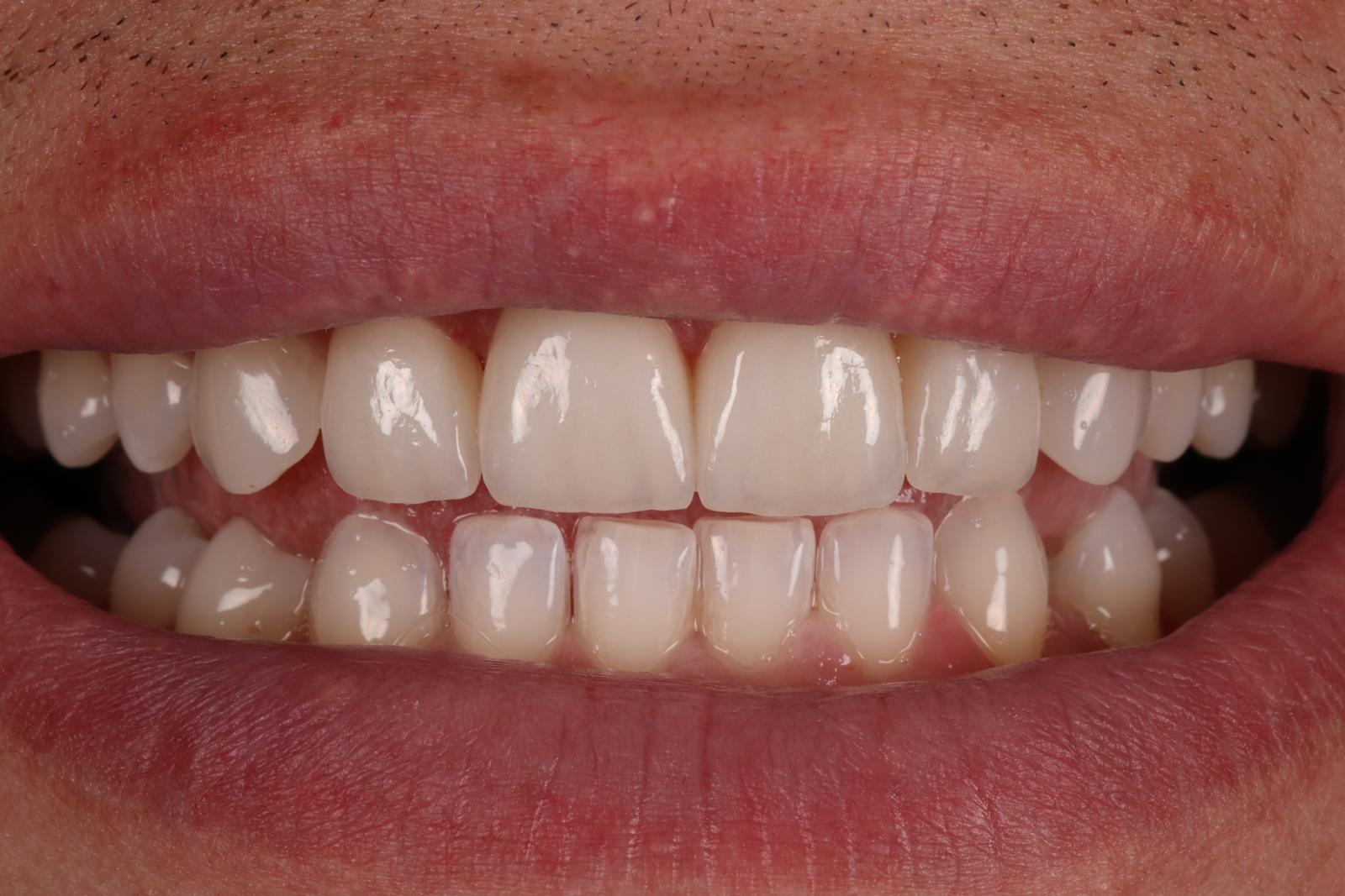
I finished my braces, but my teeth do not meet properly. Why?
Orthodontics aligns the position of the teeth. How they meet also depends on their shape and size, and sometimes these two things do not fit together perfectly.
At the end of treatment, contacts can be few or point-like, and they can settle in the following weeks. Differences in shape (small, worn or peg-shaped teeth) are not corrected by braces. If the problem is one of position, the orthodontist can make finishing adjustments, so the first conversation should be with them. Restorations do not move teeth and do not replace orthodontic finishing.
Why do gaps between the upper and lower teeth need to be addressed?
For effective chewing, teeth need stable, well-distributed contacts when you close your mouth. If some back teeth do not meet, they do not contribute properly to supporting the bite, and forces may be concentrated on fewer teeth and their restorations.
During rehabilitation, where indicated, we restore tooth shape and height so that more teeth contribute to support and chewing forces are distributed favourably. The aim is sufficient, stable, well-distributed contacts, rather than simply the highest possible number of contact points. We check contacts both when you close your mouth and during jaw movements.
We first establish why the contacts are missing. The plan may involve orthodontic finishing, restoring tooth shape or a combination of treatments; restorations do not replace a necessary orthodontic correction.
Why do gaps remain after orthodontic treatment?
The most common causes are teeth smaller than the space available, relapse, meaning the tendency of teeth to drift back, and the anatomy of the surrounding tissues.
- Teeth smaller than the space. Small or peg-shaped lateral incisors leave gaps that braces cannot close without affecting other positions.
- Relapse. If the retainer is not worn as instructed, teeth can drift back, including the gaps.
- The labial frenum or gum tissue. Sometimes it contributes to a gap returning between the central incisors.
- A gap left on purpose. The orthodontist may leave space so the teeth can then be restored to the right proportions. This is a planning decision, not a mistake.
How residual gaps are closed
Drag the slider to add material to the small teeth. The dashed line shows the target shape set on the wax-up.
- Natural tooth — fixed position
- Added composite — highlighted in green
The lateral incisors are smaller than the space available, so gaps remain on both sides.
Educational model, not to clinical scale. The percentage shows demonstration progress, not a patient’s measured gaps. Clinical composite is shade-matched to the tooth.
Build up the chewing surfaces
Move each slider to add material to a posterior tooth.
- Natural tooth
- Added material
- Dashed line: illustrative contour
A more detailed article is Why do gaps remain after braces?
Restorative rehabilitation: stages and visits
After orthodontic treatment, tooth position may be appropriate but worn or eroded teeth, differences in shape and proportions, gaps, underdeveloped cusps or incomplete occlusal and interproximal contacts may remain.
- Planning
Tooth shape, contacts and occlusion are assessed. Photographs, intraoral scanning and a wax-up help establish the final morphology.
- Restorative rehabilitation
Teeth requiring changes are rebuilt mainly with minimally invasive adhesive composite techniques. Shape, cusps, marginal ridges, interproximal and occlusal contacts can be restored. Temporary restorations, crowns, onlays or other indirect restorations may also be included when needed.
- Completion and review
Contacts and occlusion are checked and adjusted, final photographs and scans are taken, and the retainer is remade when necessary to fit the new situation.
Adhesive procedures are carried out under isolation and, when the situation allows, with magnification.
For international patients
Predominantly restorative post-orthodontic rehabilitation may require approximately 3–4 clinical visits. In many cases, treatment can be grouped into one trip of around 4–7 days, with a subsequent review if needed. Timing depends on the patient, the number of teeth treated and case complexity.
Post-orthodontic restorative rehabilitation
Photographs digitally retouched for lighting and clarity. Documentation shows an individual case; results vary between patients.
Sources
- Aschheim KW (ed.). Esthetic Dentistry: A Clinical Approach to Techniques and Materials. 3rd ed. Mosby, 2015.
- Layton DM, Walton TR. An up to 16-year prospective study of 304 porcelain veneers. Int J Prosthodont, 2007.
The information on this page is general and educational. It does not replace a medical consultation, and a treatment recommendation can only be made after examining each patient. Results differ from one case to another.
Start with a consultation and a plan
Every treatment begins with a diagnosis: clinical examination, photographs, radiographs and, where relevant, an occlusal analysis. Only then do we discuss options.